[HIDE]
There are various types of primary bone cancers depending on the type of cells of their origin. The most common primary bone cancer is osteosarcoma (osteogenic sarcoma) that arises from the osteoid tissue and usually affects long bones of the arms & the legs. The second most common primary bone cancer is chondrosarcoma that arises from the cartilage. The third most common primary bone cancer is Ewing's sarcoma that arises from the immature nerve tissue in the bones. Other primary cancers of the bone include fibrosarcoma that arises from connective tissue within the bone marrow cavity, malignant giant cell tumour that arises from the connective tissue of the bone marrow and chordoma that arises from cellular remnants of the foetal spinal cord. The exact cause of primary bone cancer is not fully understood but the researchers believe that it is caused by over-activity of the bone cells. Studies have shown that there is a much higher risk of bone cancer in those persons, who had taken radiotherapy or chemotherapy with alkylating anticancer drugs during their childhood.
Pain and swelling are the most common symptoms of bone cancer. The tumours located in or near a joint may lead to stiffness and tenderness of the affected joint, whereas the tumours in the pelvic bones and lower part of the spine may affect the normal functioning of the bladder and the bowel. Bone cancer usually leads to pathological fractures of the affected bones. Procedures used in diagnosis and evaluation of the bone cancer include a blood test, X-rays, bone scan, CT scan, fluoroscopy and biopsy.
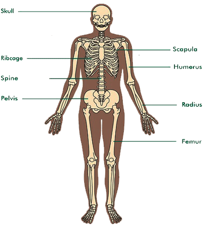
Osteosarcoma (osteogenic sarcoma) is the most common and highly malignant primary bone tumour. It is more common in men as compared to women and usually occurs during the second and third decades of life. Osteosarcoma usually affects long bones such as the humerus, distal part of the femur and proximal part of the tibia & the fibula. The exact cause of osteosarcoma is not fully understood but radiation is considered as one of the major risk factors. Histopathologically, the tumour is composed of pleomorphic cells that invade and destroy the bone. Osteosarcoma is divided into three types, i.e. the parosteal osteosarcoma, multifocal osteosarcoma and the soft tissue sarcoma.
Osteosarcoma presents as a painful bony swelling usually around the shoulder or the knee. If untreated, the tumour may fungate to form an ulcer. The malignant cells of osteosarcoma often produce an abnormal irregular bony structure. The expanding tumour forming a new bone in the soft tissue may appear as Codman's triangle and Sun-ray spicules in the X-ray films. Osteosarcoma may cause pathological fracture. The tumour usually metastasises to the lungs. Staging of the osteosarcoma is done as follows: In local stage of the osteosarcoma, the tumour is localised in the affected bone. In metastatic stage, the osteosarcoma spreads to other parts of the body. Procedures used in diagnosis of the osteosarcoma include X-rays, CT scan, fluoroscopy, bone scan and biopsy.
Chondrosarcoma is the second most common cancer of the bones. It may arise from the cartilage and the bone-forming tissue. Chondrosarcoma usually occurs during 40 to 60 years of age. It may affect the ribs, ilium, scapula and proximal ends of the femur & the humerus. Any pre-existing cartilaginous tumour may also get transformed into the chondrosarcoma. Chondrosarcoma is of two types, i.e. the low-grade chondrosarcoma (the slow growing tumour) and high-grade chondrosarcoma (the fast growing tumour). The lesions of chondrosarcoma usually have ill-defined margins and these may lead to destruction of the bone. The most common symptom of chondrosarcoma is a painful lump, usually found in the pelvic area. Chondrosarcoma often invades the adjacent soft tissues. The tumour may attain a considerable size in the flat bones. In the spine, the chondrosarcoma usually presents as a small tumour that may lead to the vertebral collapse and compression of the spinal cord. It usually metastasises to the lungs. Procedures used in diagnosis of the chondrosarcoma include X-rays, CT scan and biopsy.
Fibrosarcoma may arise from the soft tissue and the bone. Fibrosarcoma of the soft tissue is more common in men as compared to women and occurs during 40 to 50 years of age usually arising from fibrous tissue of the arms, legs and the trunk. Fibrosarcoma of the bone often occurs during 30 to 40 years of age and affects bones of the hip, pelvis, legs and the arms. Fibrosarcoma of the bone is further divided into two types, i.e. endosteal fibrosarcoma and periosteal fibrosarcoma. Endosteal fibrosarcoma causes bone destruction leading to pathological fracture and usually metastasises to the regional lymph nodes & the lungs. Periosteal fibrosarcoma invades the bone but does not metastasise to distant parts of the body. The diagnosis of fibrosarcoma is usually done by biopsy.
Synovial sarcoma is a highly malignant tumour that may arise from the muscle, connective tissue and the subcutaneous tissue. It usually occurs during 20 to 40 years of age. Synovial sarcoma often presents as a soft tissue mass in a major joint such as the knee, ankle and the wrist. It usually metastasises to the regional lymph nodes.
Malignant giant cell tumour usually originates in the epiphyseal region of long bones such as distal part of the femur and proximal part of the tibia & fibula. It is more common in women as compared to men and usually occurs during 30 to 50 years of age. Malignant giant cell tumour often presents as a swelling in the bone and the adjacent joint along with pain. There may be destruction of the bone that gives typical soap bubble appearance in the X-ray film. The growing tumour expands the bone resulting into thinning of the bone. Pathological fracture is a common symptom of the malignant giant cell tumour. Invasion of the tumour into the adjacent tissues occurs after the fracture. Malignant giant cell tumour usually metastasises to the lungs. Recurrence of the tumour is also common.
Secondary tumours of the bone are quite common. About two third secondary tumours of the bone metastasise from primary cancers of the breast, lung and the prostate; about one-sixth metastasise from primary cancers of the bronchus, kidney and the thyroid; and in the remaining one-sixth secondary tumours of the bone, the primary site is unknown. Secondary tumour of the bone usually presents with debilitating pain, impaired walking and severe neurological impairment. The destructive lesions in the bone may lead to the pathological fracture. Procedures used to diagnose secondary tumours of the bone include X-rays, bone scan, CT scan and MRI.

Disclaimer:
This content is for information and educational purposes only and should not be perceived as medical advice. Please consult a certified medical or healthcare professional before making any decision regarding your health using the content above.
Click here to go back to the list of all Articles


Bone Cancer